Gain of keratinized tissue
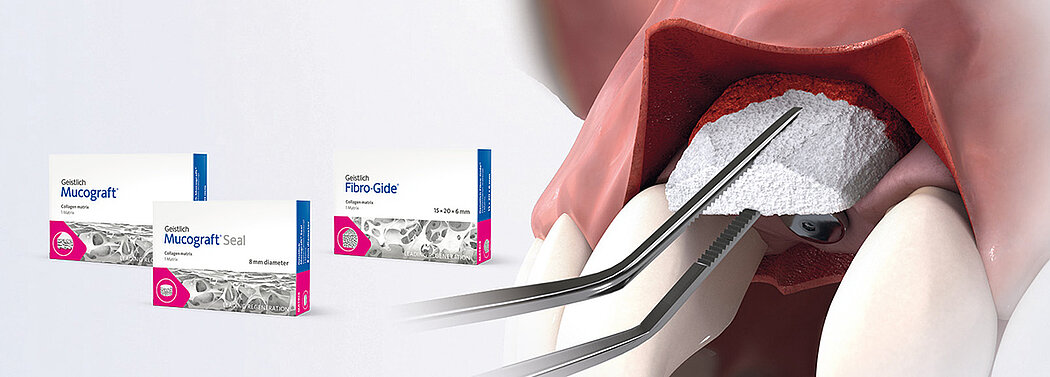
The band of keratinized tissue around teeth and implants is considered to be important in maintaining functionality and esthetics. It also enables patients to maintain good oral hygiene without irritation or discomfort.1,2
The width of the keratinized mucosa around an implant might also influence the risk of peri-implantitis.3 This remains a controversial issue.4
Autogenous soft tissue grafts, such as free gingival graft or connective tissue grafts, have proven successful in creating additional keratinized tissue around teeth and implants.5 However, harvesting those grafts, usually from the palate, is painful, technically demanding, time consuming and may lead to complications such as bleeding, pain, swelling, and occasionally also numbness or infections.6-9
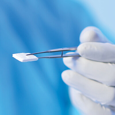
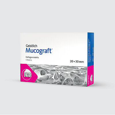
Several studies have shown successful outcomes with Geistlich Mucograft®. The collagen matrix creates the same amount of keratinized tissue as connective tissue grafts10 or free gingival grafts5,11 and helps to regenerate a keratinized tissue that resembles the surrounding native gingiva.12 Compared to autologous grafts the matrix produces a better color and texture match with the surrounding tissue that remains stable in the long term.13
References:
- Schrott AR, et al.: Clin Oral Implants Res 2009; 20(10): 1170-77. (clinical study)
- Chung DMT, et al.: J Periodontol 2006; 77(8): 1410-20. (clinical study)
- Schwarz F, et al.: J Periodontol 2018; 89 Suppl 1: S267-S290. (review)
- Greenstein G, Cavallaro J: Compend Contin Educ Dent 2011; 32(8): 24-31. (review)
- Thoma DS, et al.: Clin Oral Investig 2018; 22(5): 2111-19. (clinical study)
- Griffin TJ, et al.: J Periodontol 2006; 77: 2070-79. (clinical study)
- Soileau KM, et al.: J Periodontol 2006; 77: 1267-73. (clinical study)
- Zucchelli G, et al.: J Clin Periodontol 2010; 37: 728-38. (clinical study)
- Cairo F, et al.: J Clin Periodontol 2012; 39: 760-68. (clinical study)
- Lorenzo R, et al.: Clin Oral Implants Res. 2012; 23(3): 316-24. (clinical study)
- Nevins M, et al.: Int J Periodontics Restorative Dent 2011; 31(4): 367-73. (clinical study)
- Schmitt CM, et al.: J Periodontol 2013; 84: 914-23. (clinical study)
- Schmitt CM, et al.: Clin Oral Implants Res 2016; 27(11): e125-e133. (clinical study)
Clinical Cases
Gain of keratinised tissue
Clinical challenge
Gain of keratinised tissue around teeth
Aim / Approach
Gain of keratinised tissue in the anterior-inferior region.
Conclusion
In some cases the absence of attached gingiva is related to discomfort during brushing, persistent gingival inflammation and muscle pulling. In this case, Geistlich Mucograft® was used with the aim of gaining keratinised tissue in the buccal aspect of two lower central incisors, avoiding the harvesting of a free gingival graft from the palate. The final outcome, 6 months after surgery, shows a nice band of keratinised tissue with good colour and texture match. The result of the procedure met the patient’s expectations as brushing can now be properly executed without any discomfort. No attempt was performed to cover the exposed roots at this stage; however, the current clinical situation is now favourable, if a second surgery for root coverage is desired.
Gain of keratinised tissue
Clinical challenge
Augmentation of width of keratinised tissue around prosthetic restoration
Aim / Approach
Augmentation of the width of keratinised tissue around prosthetic restoration, avoiding the patient morbidity caused by autogenous soft-tissue grafts.
Conclusion
Mucograft® (prototype) is as effective and predictable as connective tissue graft (CTG) to gain an adequate width of keratinised tissue. The 3D-matrix shows excellent handling properties and can be used successfully in an open healing situation, reducing significantly patient morbidity and surgery time compared to CTG.
Gain of keratinised tissue
Clinical challenge
Increase of width of keratinised tissue around implants
Aim / Approach
Increasing the width of keratinised tissue around implants with Geistlich Mucograft®, while also achieving vestibule creation and oral hygiene access improvement.
Conclusion
Geistlich Mucograft® can be used as an alternative to significantly increase the zone of keratinised and attached tissue around existing implants. In addition, good texture and colour match to surrounding native tissues was observed on the mucogingival tissues regenerated with the 3D-collagen matrix.
Gain of keratinised tissue
Clinical challenge
Gain of keratinised tissue around teeth
Aim / Approach
Increasing the width of keratinised tissue without harvest of autogenous soft-tissue graft.
Conclusion
The 3D-matrix Geistlich Mucograft® may be used successfully to increase keratinised tissue around teeth without the need of harvesting free gingival graft from the palate. The aesthetic outcome is optimal and stable over time (1 year).
Gain of keratinised tissue
Clinical challenge
Widening of attached gingiva prior to implant placement
Aim / Approach
Widening of the attached gingiva using Geistlich Mucograft® for complex implant rehabilitation prior to augmentation and implant placement.
Conclusion
The use of Geistlich Mucograft® for widening of the attached gingiva shows a good increase of width around teeth and implants comparable with autologous grafts – with significantly reduced morbidity by avoiding the palate wound. The shrinkage of the xenogeneic collagen matrix is higher than that of a free gingival graft (FGG), so an overextension of the preparation and matrix is mandatory. The colour match is excellent and much better than with a FGG.


